Managing A Calf Tear
Calf tears, know in the medical world as Gastrocnemius tears, are a commonly torn lower limb muscle accounting for 12% of all muscle tears (Armfield et al 2006). They occur frequently in athletic populations with research suggesting that the medial head of the gastrocnemius is the third most commonly strained muscle in elite athletes. It is also commonly injured in weekend warriors playing recreational sport and the general population after sudden movements like chasing after the dog, running after the bus or dashing across the road.
Anatomy
The gastrocnemius is one of the main plantar flexors of the ankle effectively allowing you to point your foot down to “push off”. It is comprised of two muscle bellies, with the medial head having a mechanical advantage. It is therefore able to generate more force, making it more susceptible to injury compared to the lateral head. “Two-thirds of calf injuries occur at the junction of the fascia between the medial head of the gastrocnemius and the soleus (the other big calf muscle). Injuries to the lateral head of the gastrocnemius occur in up to 14% of patients in some case series, but injury may occur anywhere from the proximal origin to the mid-belly to the fascial junction with the soleus.” (Bright et al 2017)

How is it injured?
Muscle tears commonly occur when force placed through the muscle outweighs its capacity and the gastrocnemius is no different. This can result during falls, tackles or explosive movements placing high demands on the calf muscle. Other factors such as fatigue, technique and biomechanics can increase the likelihood of the muscle becoming overloaded and failing under force.
How are symptoms described?
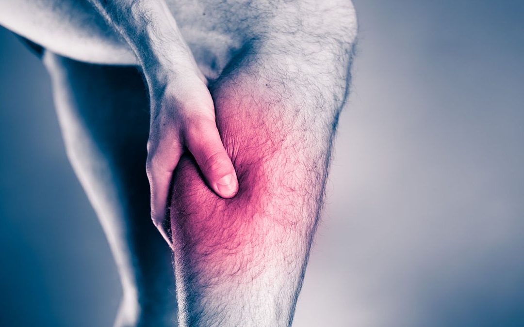
Similar to the history of an Achilles tendon rupture, people often think they’ve just been hit in the leg by a ball or other object and potentially hear a “pop” or “snap”. In nearly all cases, they will feel a sudden pain in the back of their calf particularly at the muscular tendinous junction. If the Soleus muscle is damaged pain might be incurred lower in the leg and when contracting the muscle against resistance with the knee bent.

Diagnosis & Examination
Physiotherapists are highly qualified professionals with expert knowledge and skills in the diagnosis of muscle injuries. A thorough subjective and objective examination performed by a Physiotherapist is sufficient to diagnose a calf tear. Early diagnosis is key to ensuring that initial management is tailored to the patient and specific structure that is injured.
A thorough examination will reveal the following signs: usually a patient will walk into the clinic with a limp, and depending on the severity of the tear, they may be unable to weight bear on the injured leg at all. In higher grade tears there may be a palpable gap that we can feel over the site of the tear and depending on how acutely they present, there may be associated bruising and discolouration of the skin. The injured patient will also lose range of motion in in their ankle. Typically, they will be unable to raise on their toes performing a double or single leg calf raise however this also depends on the severity and grade of the tear.
“The vast majority of calf injuries can be diagnosed and managed without any additional imaging” (Bright et al 2017). Occasionally an ultrasound can be used to determine the size of the tear however they are not always accurate, and although clinically a tear has been diagnosed sonographers can miss these from time to time so ultrasounds are not routinely requested as it can lead to misdiagnosis. Further imaging such as MRIs are rarely utilised unless differential diagnoses are suspected and need to be rules out.

Treatment and Evidence
Initial stages of treatment aim to reduce pain, swelling and further injury. Crutches may be necessary for patients that are unable to weight bear. They should only be used for a short amount of time as early mobilisation allows muscle fibres to regenerate and organise more effectively (Jarvinen TA et al. 2005). Within 10 days, scar tissue that has formed over the tear will have the same tensile strength as normal muscle and gradual loading can begin (Dixon et al 2009).
Ice is useful in the acute stage and patients will often benefit from the use of Tubigrip compression to support the injury site.
In the early stages as symptoms improve it is important to restore normal walking patterns to avoid developing compensations. Our physiotherapists will help our patients activate and use the correct muscles with exercises and gait retraining. Incorporating an exercise program to improve range of motion, strength, endurance and motor control is essential for recovery and should be implemented once symptoms allow. Gentle stretching is important to lengthen the muscle, improving elasticity and distending the scar tissue appropriately (Jarvinen et al 2005). Strengthening exercises will be provided for the muscles in the calf and foot progressing in difficulty until the injured leg’s strength and endurance is comparable to the non-injured leg. Brukner and Khan recommend addressing biomechanical factors that preceded the injury such as muscle tightness, lumbopelvic dysfunction or knee/ankle related abnormalities. Proprioception exercises (a form of balance retraining) must also be utilised as there is a loss of balance and control associated with calf tears whilst dynamic sport specific exercises are implemented in preparation for returning to full activities. Completing rehabilitation is the most effective way to return to performance/activity and prevent recurrent or secondary injuries.
ARE YOU STRUGGLING WITH CALF PAIN OR A MUSCLE STRAIN/TEAR?
GET AN ASSESSMENT & TREATMENT WITH OUR AMAZING TEAM OF PHYSIO’S.

- Get crystal clear about what’s causing your Pain – Get An Accurate Diagnosis
- Find out the most important steps for you to get pain free fast!
- Understand how to manage your load and activities without suffering the consequences of further injury
- Find out the simple yet essential exercises that can give you relief and speed up your recovery
- Leave the session with the confidence of knowing exactly what to do to get symptom free and get back to the things you love