Lower Back Injury
Has your child been complaining of lower back pain or stiffness during sport and activity?
This might be a helpful article to read as we will give you some insight into an unfortunate common cause of adolescent back pain known in the medical world as Lumbar Spondylolysis.
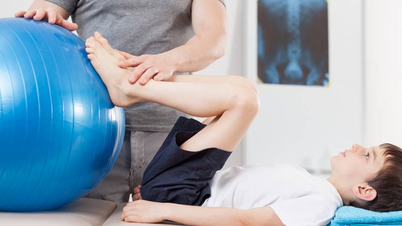
Lumbar spondylolysis is an injury to the pars interarticularis of the lumbar spine in the lower back region. The term includes a spectrum of bone stress injuries (stress reaction and stress fractures) and defects (non-united fractures). It has a relatively small incidence in the general population of 4-9%, however, it is far more common in the adolescent athletic population, accounting for about 50% of all low back pain in this category (McTimoney et al, 2003). Sports that involve repetitive lumbar extension, such as gymnasts, tennis players, cricket bowlers and dancers, are most at risk. It will generally present as pain localised to the lumbar spine with symptoms progressing in severity with continued activity. When identified most patients with an injury to the pars respond well to conservative management, including core strengthening, postural control, physical therapy, and hydrotherapy (Mobbs, R. J. et al. 2019).
Anatomy
The pars interarticularis is part of the vertebra located between the inferior and superior articular processes of the facet joints. The adjoining lamina and pedicle have also been shown to be involved in the bone stress injury. This posterior region of the spine is loaded through repetitive trunk extension, rotation and side flexion movements leading to bone stress. These stress reactions and fractures can occur on one or both sides of the vertebrae.

How is it injured?
Lumbar spondylolysis is caused due to a repetitive mechanical stress of the lumbar spine leading to bone stress reaction and eventual fracture. Less commonly, a single traumatic event or stressor may also lead to a pars defect. Sometimes children are born with a very thin pars area which puts them at increased risk for this injury. Research supports that there are hereditary and acquired risk factors that can make one more susceptible to the defect. In a young athlete, the spine is still growing which means there are many ossification centres, leaving points of weakness in the spine. This leaves young athletes at increased risk, particularly when involved in repetitive hyperextension and rotation across the lumbar spine.
How are symptoms described?
Patients will typically describe a gradual onset of lower back pain, usually associated with an increase in training load or repeated loading of the spine. Spondylolysis may initially present asymptomatically with patients describing stiffness only but can progress quickly to pain and dysfunction with continued activity. Symptoms are normally reproduced with lower back side bending, twisting and leaning backwards, with pain experienced in the region of the lower back with possible referral to the gluteal area and back of the thigh. Local back muscles may exhibit tenderness and spasm which can assist in determining which segment of the spine is involved.

Diagnosis & Examination
When a patient presents with symptoms, there are general signs and symptoms a clinician will look for:
- Clinical Signs:
- Pain on completion of the single legged lumbar extension test
- Pain on combined extension, rotation and lateral flexion movements
- Excessive lordotic posture (increased “arch” in lower back)
- Tenderness on palpation of the lower back and muscle spasm
- Biomechanical deficits often include poor gluteal stability and core control on testing
- Symptoms:
- Unilateral (one sided) low back pain
- Pain that radiates into the buttocks or legs
- Onset of pain can be acute or gradual
- Pain that can restrict daily activities
- Pain that worsens after strenuous activity
- Pain aggravated with lumbar hyperextension (excessive leaning backwards)
- An increase in load during specific activities or sports
Radiological imaging has a great importance in diagnosis. When suspicious of spondylolysis, a fracture may be seen on an oblique x-ray and CT scans can confirm and grade the severity of the injury. MRI tests can pick up bone oedema (swelling) in cases where there is no pain but bone stress is present and they can also help diagnose symptomatic stress reactions, as well as stress fractures and non-united defects, however CT and SPECT can provide more detail and staging of a fracture (Brukner and Khan, 2017). Your physio and treating doctor will arrange the appropriate scans and referrals needed.

Differential Diagnosis
It is important to conduct a thorough physical assessment and imaging if indicated to exclude other pathologies that may present similar to spondylosis as differential diagnosis can alter treatment and also affect a patient’s prognosis.
Treatment and Evidence
Young patients with spondylolysis generally receive conservative management as their initial treatment. Conservative management can produce excellent clinical and radiological outcomes, (particularly in young athletic populations) however return to sport can take between 3 to 12 months. Most patients do not require surgery but, if symptoms are not relieved with non-surgical treatments, or when the condition progresses to high grade spondylolisthesis (stress fractures on both sides), then patients may require surgery.
Conservative treatment of a pars defect can be broken down into 3 primary phases as described by Brukner and Khan, 2017.
0-8 weeks: Fracture healing phase
The aim is to protect the area optimising bone healing and preventing further progression. Activities that load the vertebra should be ceased (running and jumping etc), as well as strength training involving repetitive loading or end range movement. A tailored rehabilitation program should be commenced in consultation with a physiotherapist to target safe strengthening, muscle control/activation, and flexibility of the lumbopelvic region. Rehabilitation exercises in this phase aim to keep overall muscle strength in a safe manner and also address kinetic chain and lumbo-pelvic deficiencies (often arising as gluteal and core dysfunction). Cardiovascular exercise such as walking, or cycling are usually safe to continue initially. The use of a thoracolumbar brace can also be considered during this phase.
9-16 weeks: Protected Reloading Phase.
In this phase, pain and clinical signs on testing should be absent during Activities of Daily Living (ADLs) and also during rehabilitation exercises. With guidance, low impact exercise should be introduced to start loading the spine to stimulate bone healing. Rehabilitation exercises should be progressed functionally, incorporating loaded strengthening (dumbbell squats/lunges), and body weight extension/rotation exercises. Around 12 weeks, jogging may be able to be progressed to higher intensity running, and toward the end of this phase dynamic and sports specific training can be incorporated proving the patient meets certain test requirements. It is important that progression of activity remains pain free with no symptoms on clinical testing. It is also very important to also assess technique of specific sports/activities. An understanding the biomechanical demands of different tasks can be a really important aspect of altering the load and stress on the lumbar spine. Communication between physio’s and coaches can be an important factor in this regard.
17 weeks onward: Transition to Full Function and Sport
Increases in rehab and training load, and transition to sport/unrestricted exercise including plyometric training should be gradually progressed, with careful monitoring of symptoms. Any aggravation of symptoms or increased signs on clinical testing is a sign that activity needs to be regressed or ceased. Unfortunately, many patients neglect this monitoring phases of rehabilitation as return to activity has occurred already, however careful monitoring needs to take place.
Radiological evidence of healing would be expected at this stage, with resolution of bone oedema at 16 weeks on MRI coinciding with bone healing on CT scan.
In the case of symptomatic pars defect in adolescent patients, roughly 75% to 95% will improve with appropriate conservative management (Mansfield et al, 2019).
Overall the diagnosis and management of pars interarticularis injury or spondylolysis is extremely important as it is frequently misdiagnosed and can potentially affect clinical outcomes, activity levels and patient satisfaction. If you require any further information, please don’t hesitate to contact our clinic.

ARE YOU OR YOUR CHILD TROUBLED BY BACK PAIN?
GET A COMPREHENSIVE ASSESSMENT & TREATMENT WITH OUR TEAM OF AMAZING PHYSIO’S.

- Get crystal clear about what’s causing your Back Pain.
- Find out the most important steps for you to get pain free fast!
- Understand how to manage your normal activities without suffering the consequences of pain
- Find out the simple yet essential exercises that can help you get lasting and fast relief
- Leave the session with the confidence of knowing exactly what to do to get symptom free and back to living life without pain