The medial and lateral menisci are two C-shaped fibrocartilaginous structures located on the top of the tibia. They act as a shock absorber, stabiliser and are an attachment site for tendons, muscles and ligaments.

Meniscal tears can be:
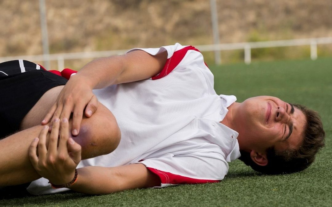
Acute: A result of weight bearing and twisting on the leg. These commonly occur in sport, and can be associated with ACL tears.
Degenerative: Occur over a long time with repetitive weight bearing activities (such as kneeling & squatting) and progress with age.

Signs & Symptoms
- Swelling (Commonly delayed after injury)
- Restricted knee movement (Can include ‘locking’ sensation)
- Pain with weight bearing

Risk Factors
- Involvement in sports requiring running, jumping or pivoting
- Previous knee injury (particularly ACL rupture)
- > 60 years of age
- Occupational activities: Frequent squatting, kneeling or stair climbing
- Poor biomechanics – including lower kinetic chain dysfunction

Diagnosis/Assessment
- Reduce knee joint range of motion
- Inability to fully straighten knee
- Positive McMurray’s test (Pain +/- click or clunk
- Tibiofemoral joint line ternderness
- Knee joint swelling/effusion present
- Inability to squat or “duck walk” MRI can assist along side clinical diagnosis
Meniscus injuries are the second most common knee injury, with a prevalence of 12% to 14% of all knee injuries.
Conservative approach
- Symptoms develop 24 – 48 hours post injury
- Minimal injury or unable to recall injury
- Able to weight bear
- Minimal swelling
- Full ROM with pain only at end of range
- Pain on McMurray’s test at end of range flexion only
- Previous history of rapid recovery from similar injury
- Early degenerative changes on x-ray
Surgical Approach
- Severe twisting injury, unable to continue playing
- Locked knee or severely restricted ROM
- Positive McMurray’s test- “clunk”
- Pain on early range flexion on McMurray’s test
- Presence of ACL tear
- Little improvement after 3-4 weeks of conservative treatment
- Type, location, severity or size of tear
What Does The Evidence Say?
Herrlin (2017) found when comparing degenerative meniscus tears, patients that had arthroscopic surgery plus an exercise program had no additional benefit to an exercise program alone. This study validates that an exercise program compiled by a physiotherapist can be as effective as surgery.
Brukner (2012) suggests, a physiotherapy approach should be undertaken for 3-4 weeks following meniscal tears, however failure with conservative management would require surgical intervention.
Why is physio important?
Meniscal injuries can cause accelerated wearing of cartilage and lead to early onset of osteoarthritis in the knee (Jarraya et al, 2017). Therefore management should be sought in the early stages to reduce the pressure on the meniscus as well as secondary compensations.
Meniscal tear patients with higher levels of leg strength had less pain, less difficulty completing daily tasks and had improved mobility compared to those with reduced strength (Luc-Harkey et al, 2018). This shows the importance of a strengthening program to address the supporting muscles around the meniscus.
Early physio management after meniscal injury helps to normalise the injured persons gait and improve muscle activation patterns, improving their chance of non-surgical management. This typically leads to less pain, less dysfunction and a better outcome with an earlier return to ADL’s, sport and normal function.
GET AN ASSESSMENT & TREATMENT WITH OUR AMAZING TEAM OF PHYSIO’S.
- Get crystal clear about what’s causing your injury or pain
- Understand what activities you can do without suffering consequences of pain or instability
- Find out the simple yet essential exercises that can help you achieve amazing result
- Leave the session with the confidence of knowing exactly what to do to get symptom free and back to living life to the full